- Mon - Sun : 24 Hours Open
- +91 8058050454
- info@galaxyimaging.in
- Vashi Navi Mumbai 400703
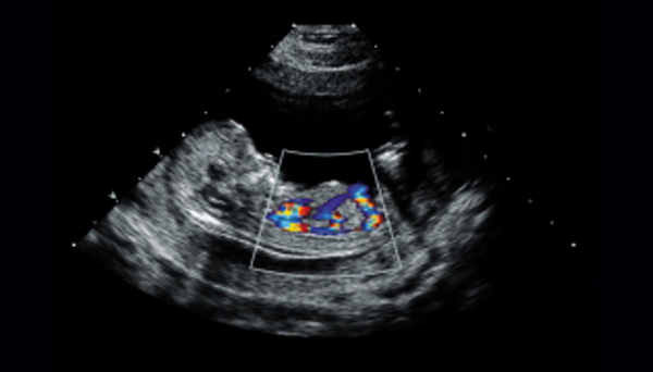
Fetal echocardiography is a specialized ultrasound test that evaluates the structure and function of a baby's heart while still in the womb.
It is typically performed between 18 and 24 weeks of pregnancy to detect congenital heart defects and assess the baby's cardiac health. The test uses sound waves to create detailed images of the fetal heart, helping physicians diagnose and plan treatment for any heart abnormalities before birth.
Uses ultrasound technology, which is safe for both mother and baby with no radiation involved